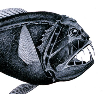
The Paper:
Dunlap, P. V, Takami, M., Wakatsuki, S., Hendry, T. A., Sezaki, K., & Fukui, A. (2014). Inception of bioluminescent symbiosis in early developmental stages of the deep-sea fish , Coelorinchus kishinouyei ( Gadiformes : Macrouridae ). Ichthyological Research, 61, 59–67.
Background:
Have you ever gone swimming in the ocean at night and noticed tiny lights when you splashed around? Those lights are produced by little organisms in the water via a chemical reaction, a process known as bioluminescence. Bioluminescence is extremely important in the ocean and is used by a variety of organisms including bacteria, jellyfish, squid, and even fish. Bioluminescence is often times used as a signaling device to locate mates, attract prey, and confuse predators. There are two ways an animal can produce bioluminescence:
- By producing the light directly via chemical reactions in their body
- By relying upon a symbiotic relationship with bioluminescent bacteria
In this symbiotic relationship, the host animal provides the bacteria with a place to live (inside the host’s body) and with necessary nutrients and oxygen to grow and reproduce. In return, the bacteria produce light that the host can then use for signaling. Before this relationship can begin, the bacteria must colonize within the host. Some bacteria may be directly transferred from parent to offspring but other hosts must acquire the bacteria directly from the environment (i.e. by eating them).

Over 460 species of fishes utilize bacteria to produce light. While these fishes can be found everywhere from coastal habitats down to the depths of the ocean, 248 of these species belong to a group of deep-sea fishes known as the rattails (Fig. 1), most of which belong to the genus Coelorinchus. Despite the abundance of rattails and their growing importance in commercial fisheries, very little is known about the bioluminescence produced by these fishes. This study investigates the structure of the luminescent gland in larval and juvenile rattails (Coelorinchus kishinouyei) to elucidate (pun intended) how and when these fish acquire their glowing companions.
Methods:
This study used bottom trawls to collect specimens in Suruga Bay in Honshu, Japan. Scientists collected rattails with trawls ranging in depth from 186 – 500 m. The rattails identified as Coelorinchus kishinouyei were sectioned (think of it like getting ham sliced for you at a deli, but instead of ham it is a fish getting sliced into thin sections of tissue) and stained with tissue dyes to be able to observe the structure of the luminescent organ. This technique allows for scientists to examine thin layers of tissue much more closely.

In order to determine the type of bacteria living in the fish, the scientists removed the fish’s luminescence glands and extracted the bacteria from the glands to grow in cultures. These bacterial cultures were then genetically sequenced and identified to determine what type of bacteria help these fish glow.
Results:
The rattails were found to have a luminescent gland in their abdomen branching off of the intestine near the anus. In smaller fish the gland was present as just a small nub, but grew to a larger bean-shaped organ in larger fish. While all fish examined had a luminescent organ, not all of them had been colonized by bacteria, as noted by empty (uncolonized) finger-like projections (Fig. 2).
Some larvae had bacteria in their luminous organs while others did not, but all juvenile light organs contained at least some bacteria (Fig. 3). As bacterial colonization occurs, it appears that there is a change in the shape of the organ with the finger like projections switching from a horizontal orientation (Fig. 2) to a vertical orientation (Fig. 3). The scientists hypothesize that the change in shape of the gland helps to better transmit the light produced by the bacteria. There are very few examples of bacteria causing the shape of an organ to change, making this a very interesting find.
Conclusions:

As in most deep-sea fish, the rattail eggs are laid in the deep ocean and float to shallower waters, where the fish will hatch. As the fish grows, it will move back to deeper water where it will settle and live as an adult. The study found that these life history changes may determine when the luminescent bacteria form colonies within the fish. It appears that the luminescent gland begins to form only after the larvae have developed substantially and begin moving back into deeper water. By the time the larvae reach the deep-sea where they will settle, the luminescent gland is fully formed, but does not yet contain bacteria.
The researchers hypothesize that the larvae encounter the bacteria and ingest them with seawater, or zooplankton prey. The bacteria travel through the gut until they reach the beginning of the light organ, which branches off of the small intestine. The bacteria then travel into the luminescent organ where they form colonies and cause the organ to change its shape. The researchers also hypothesize that larval rattails may benefit from settling in an area where adult rattails are present. As the adult luminescent organs become full with bacteria, extra bacteria are released into the surrounding environment, thereby increasing the amount of bacteria in the water that can be found by newly settling larvae.
Significance:
The mode by which deep-sea fishes acquire luminescent bacteria is poorly understood. While more studies are needed to test the hypotheses posed in this article the study proposes an interesting mechanism for bacterial acquisition relating to changes in the life history of deep-sea fishes. Previous work has been unable to focus on life history stages in deep-sea rattails. As these fish become increasingly important in fisheries, more information will be needed on their life history and biology. Thanks to technological advances in the field, this study has laid the groundwork for future studies but also highlights the need for scientific exploration in the poorly understood world of the deep sea.
I received my Master’s degree from the University of Rhode Island where I studied the sensory biology of deep-sea fishes. I am fascinated by the amazing animals living in our oceans and love exploring their habitats in any way I can, whether it is by SCUBA diving in coral reefs or using a Remotely Operated Vehicle to see the deepest parts of our oceans.


2 thoughts on “Deep-sea Lights”